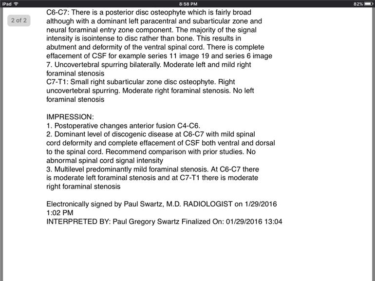
Hi! Does anyone know what this means?? I appreciate any help to understand.....
MRI Results Cervical: Hi! Does anyone know what this... - NRAS
MRI Results Cervical

You probably know that there are 7 vertebrae in your neck, and that C7 is at the bottom, running up to C1 at the base of your skull.
So it is explaining the changes to the vertebrae seen on the MRI, and that you have most problems with the two that are lowest down.
Oesteophyte means you have a bit of extra bone that has grown, and foraminal stenosis means that your spinal cord inside the vertebrae is getting squished. Effacement of CSF means that something is impinging on the spinal cord, and isointense means that it is showing up brighter on the MRI than it should. So basically the bits between your 6th and 7th cervical vertebrae, and the 7th and the first one at the top of your back have problems. Which you probably know as you're in pain!
Presumably you have had some surgery on your neck? So that is the reference to the 4th, 5th and 6th vertebrae.
Morning TerriP7. It's always advised that any reports (especially the ones you don't understand) are explained to you by a medical professional, your GP or PC will know exactly what it means & are best placed to show you, maybe with the help of a model of the spine, just what is going on.
Whilst we have an idea what the report says we're not medically qualified so it's advisable to make an appointment with your primary carer or better still your Consultant (or whoever ordered the MRI) to discuss the findings. This way any concerns can be raised directly with him/her & any questions answered.
I hope you're not in too much pain, I know back pain is difficult to live with daily. 
Hi
Interpreting the MR results is straight forward for those of us in the Radiology field as the previous reply has done so well . The most important thing we need to know as a patient is how does this impact on me , what is the treatment or way forward and what is my prognosis in the long term? All of these questions can only be answered by the consultant in charge of your care. Try not to worry about the words and what they mean( easier said than done , I know) . This report is only 1 piece of the whole jigsaw and the person in charge of your care will have all the pieces of the puzzle and hopefully discuss the way forward with you at your next appointment. Hope all goes well xx
Please ask your Consultant all the above good questions. I had my appointment last week and ? results. I was not given any of these answers. My fault partly should have pressed but the Dr I saw was not helpful. I was over awed.
You have good advice by the above replies. Good luck and best wishes.
All the replies are good, especially the first which answered the question of interpretation. Also the comment on why you were sent for the MRI. I had a similar report, stenosis caused by a discs broken down (by RA) which extruded into the canal inside the vertebrae causing the cord to be crushed never mind 'squished'. In my case the cross sections in the MRI showed the cord in good order lower down like a 'black fried egg' a black circle (the nerve bundle) surrounded by a clear circle (fluid) and another ring the (outer part of the cord). But between C6 to C4 this changed into a grey blob. At C2 and C1 it was back to the 'fried egg' pattern.
Oh yes and the bone growths as well causing foraminal stenosis. Foramina are holes each side of the vertebrae through which nerves from around the body pass and merge with the spinal cord.
Nerves carry electrical impulses to and from the brain the send messages about temperature, touch and pain to the brain and they carry messages from the brain to muscles setting as many as are required to work as needed.
Stenosis damages the nerves and can prevent messages from travailing at all or reduce there signal strength. Each one has an insulating layer and if that is damaged messages can 'leak' from one nerve to another.
Now I do not want to alarm you. The comment about knowing what pain and difficulty you are experiencing is pertinent. Presumably you would not have been sent for an MRI without good cause! Go back to the doctor and make notes if you have not understood him, perhaps take someone with you who will stay focused.
In my case the consequences were severe. First I was diagnosed with RA, even though controlled by medication, I was still deteriorating. 6 months later I was sent for the MRI because of the pain in my arms and legs, I was barely able to stand or walk unaided. I had gone from normal to broken in 6 months! I had an operation 14 months ago to relieve the pressure and stabilise the vertebrae with steel posts. It was 50% successful - still in a lot of pain but I can just about walk 150 metres with a stick and stand for two minutes. The Neurologist says the nerves are scarred and it is unlikely they will recover, time will tell. ( The RA is, they say, is still well controlled by the medication. )
Without the operation I would have been double incontinent and in a wheel chair!
So get back to your doctor and discover the prognosis and options that you have.
Thank you all so very much!!
