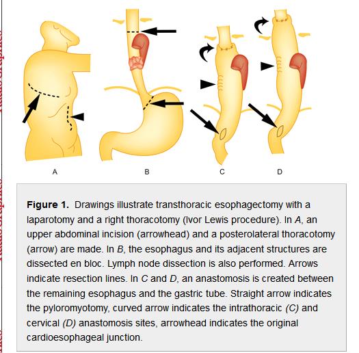
Above is a relatively simplified diagram of the principal stages of this operation.
The two major incisions are shown in A, as relates to a small to medium sized tumour in the middle to lower portion of the eosophagus. If the carcinoma involved the upper end of the eosophagus then there will probably be a third incision in the cervical area, that is on the left side of the neck. [I have had this three stage operation]
B. Indicates the area of a tumour.
C. & D. Illustrate a short pull-up and a long pull-up through the diaphragm, determined by how much of the oesophagus needed to be removed.
It is abundantly clear how the stomach itself is salvaged, cut up and fashioned into a tube to bridge the gap caused by removal of the eosophagus; approximately from navel to Adam's Apple in the worst case.
Note that the straight arrows pointing to an ellipse in C. & D. show that what had been the entry into the top of the normal stomach is now at the bottom of the reconstituted stomach tube.
Also observe how the exit bend out of the normal stomach down into the Duodenum has been straightened out by the pull-up - this impairs the natural action of the Pyloric Sphincter leading to early and late discharge (Dumping/Congestion/Pain/Pseudo-satiation etc)
Is it any wonder that our normal functions are mucked up? ( should that be an 'f' for an 'm' ?)
