Photos of my AVM from August 2012 (my surgery) and this month!
Before and After: Photos of my AVM from August 201... - Headway
Before and After
Written by

B_S_A
To view profiles and participate in discussions please or .
11 Replies
•
Hi , this is interesting, do you know how to read these things?
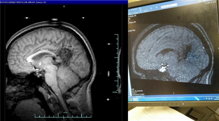
That messy looking thing in the middle of my brain is my AVM back in 2012. On the left it's pretty pronounced, but it's just sort of a black void on the right
, proof of repair, excellent. Do the white areas light up because the neurologist highlighted them, or is it how contrast dye can affect an area that is ok.. i really really dont know about this stuff my scans were never talked about. You are doing so well. r espect for working so hard on the studies.
Wow you can really see the difference.
Looks pretty neat & tidy now don't you think. ")
I remember seeing my scan at my first recall appt ; it was fascinating to see the awful mess caused by the bleed on the 'before' scan, then the 'after' scan showing the titanium coil neatly in place in a very clean looking brain.
It was an emotional experience sitting beside my 'hero' surgeon, whilst viewing the results of his skilfulness up on the screen and looking in on my own brain with hindsight.
Have a great weekend Ben. x
Good stuff : ) x Are they different types of scan ?
Hi Ben,
I don't know a lot about AVMs but I do know a thing or two anout scans. I don't like the white areas much.
You would see a long white area in my brain now as that would be my shunt but before there used to be a white, egg shaped area which represented my first tumour.
On the before scan, there is a long, curved white part. That is the AVM right? And at the base of the brain where the brain stem meets the hypathalamus (I think) is a few dim white blotchy areas. What is that?
Is that a part of the AVM?
In the recent scans the long, curved white area is gone which is good :). But at the base those white blotchy areas look a bit brighter. What does that mean?
I hope it is nothing and that is just a normal part of the brain.
Take care,
MJ
I have white patches all over my brain scans, i never saw my brain scans and nothing was ever explained to me my scans werent even mentioned. I sent for the hospktal records personally, which is how ive now seen the brain scans, and i will get the scans reviewed by a private neurologist soon. Everything is in hand so no worries. Youve made great progress Ben.
Hey Matt, sorry I don't come around as often as I used to and must have missed that.
That long curved white section is just the Corpus Callosum. Blotches on the Pons and brain stem are just small deposits of fluids and fats found in all our brains.
My AVM is just to the right of the Corpus Callosum on the scan (the darker patch towards the back of my brain).
The reason those white areas are gone in the second scan is because it's a CT scan, whereas that first scan was an MRI. CT scans don't show the resolution that an MRI, nor does it pick up these fluids or fats, which is why that curved section isn't there. The small section of white on the second scan I'm told was an anomaly of the scan itself, rather than my brain.
Basically...all good news ")
I haven't been on these Headway forums for ages now.
I see, these were scans from two different machines. Yes, the MRI scans show up more clearer.
Do/did you have an injection in your arm half the way through the MRI scan?
MRI scans sound like demented woodpeckers :).
I'm glad the white parts in the scans were nothing to worry about though and I forgot about the dark area which was the AVM :).
Take care,
MJ
Not what you're looking for?
You may also like...
Images of my Brain Before and After Aneursym Surgery
Inspired by photos on here I was just fascinated by, I thought I'd share a few photos of my brain...
Mentioned this before...? Twitching.
Hi All,
I did start a thread a few weeks back regarding "twitching" or head shacking ticking but I...
Not had this before...?
Hi All,
Not feeling very good today and iv not felt like this ever before. Feels like iv got a 5kg...
Anyone know how long it takes before you can drive after TBI?
I had a serious motorcycle accident on 3rd April 2014 and was in an induced coma for a week. I have...
How long before.....
It's been 3 months now since my craniotomy. I was told after the surgery no driving for 3 months....