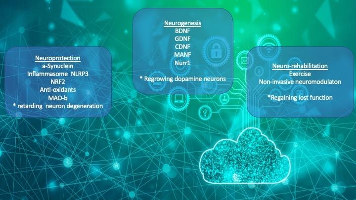
I think I've got the neuroprotection and neurorehab down pretty well. Next up, neurogenesis...
Until recently, neuroscientists believed that the central nervous system, including the brain, was incapable of neurogenesis and unable to regenerate. However, stem cells were discovered in parts of the adult brain in the 1990s, and adult neurogenesis is now accepted to be a normal process that occurs in the healthy brain. 1
Nurr1…….
Increased expression of alpha-synuclein (ASYN) and decreased expression of Nurr1 are associated with Parkinson's disease (PD) pathogenesis. These two proteins interact functionally and ASYN overexpression suppresses Nurr1 levels.
ASYN pan-neuronal overexpression coupled with Nurr1 hemizygosity followed by Nurr1 repression in aging mice results in the manifestation of a typical PD-related phenotype and pathology.
Our experiments indicate that the expression levels of ASYN and Nurr1 are critical in the dysregulation of the nigrostriatal DA system. 2
Nuclear receptor-related 1 (Nurr1) protein has been identified as an obligatory transcription factor in midbrain dopaminergic neurogenesis.
Our analysis identified ~40 NURR1 direct target genes, many of them involved in essential protein modules such as synapse formation, neuronal cell migration during brain development, and cell cycle progression and DNA replication.
Specifically, expression of genes related to synapse formation and neuronal cell migration correlated tightly with NURR1 expression, whereas cell cycle progression correlated negatively with it, precisely recapitulating midbrain dopaminergic development.
Overall, this systematic examination of NURR1-controlled regulatory networks provides important insights into this protein's biological functions in dopamine-based neurogenesis. 3
In order to understand the biological processes underlying dopaminergic neurons (DpN) regeneration in a 6-hydroxydopamine(6-OHDA)-induced adult zebrafish-based Parkinson's disease model, this study investigated the specific phases of neuroregeneration in a time-based manner.
Gene expression of nurr1 at day three, nine, 14, 18, 22 and 30 postlesion was quantified. It was found that whilst cell proliferation remained unchanged in the area of substantial DpN loss, the ventral diencephalon (vDn), there was a transient increase of cell proliferation in the olfactory bulb (OB) and telencephalon (Tel) seven days postlesion
The significant increase of EdU-ir/ TH-ir cells in the vDn 30 days postlesion indicates maturation of proliferative cells (formed between day five-seven postlesion) into Dopamine producing neurons. 4
We previously demonstrated that the pharmacological stimulation of Nurr1 by amodiaquine (AQ) promotes spatial memory by enhancing adult hippocampal neurogenesis.
This study aimed to examine changes in the cell cycle-related molecules involved in adult hippocampal neurogenesis induced by Nurr1 pharmacological stimulation. Fluorescence-activated cell sorting (FACS) analysis showed that AQ improved the progression of cell cycle from G0/G1 to S phase in a dose-dependent manner.
Our results demonstrate that the pharmacological stimulation of Nurr1 in adult brain cells promotes the cell cycle by modulating cell cycle-related molecules. 5
Previous studies have documented that orphan nuclear receptor Nurr1 plays important roles in the midbrain dopamine (DA) neuron development, differentiation, and survival.
Furthermore, it has been reported that the defects in Nurr1 are associated with Parkinson's disease (PD).
Thus, Nurr1 might be a potential therapeutic target for PD. Emerging evidence from in vitro and in vivo studies has recently demonstrated that Nurr1-activating compounds and Nurr1 gene therapy are able not only to enhance DA neurotransmission but also to protect DA neurons from cell injury induced by environmental toxin or microglia-mediated neuroinflammation. 6
Nurr1 is an orphan nuclear receptor that is essential for the differentiation and maintenance of dopaminergic neurons in the brain, and it is a therapeutic target for Parkinson's disease (PD).
During the screening for Nurr1 activators from natural sources using cell-based assay systems, a methanol extract of the combined stems and roots of Daphne genkwa was found to activate the transcriptional function of Nurr1.
Additionally, treatment with genkwa components 1 and 2 inhibited 6-hydroxydopamine (6-OHDA)-induced neuronal cell death and lipopolysaccharide (LPS)-induced neuroinflammation. Moreover, in a 6-OHDA-lesioned rat model of PD, intraperitoneal administration of 2 (0.5 mg/kg/day) for 2 weeks significantly improved behavioral deficits and reduced tyrosine hydroxylase (TH)-positive dopaminergic neuron death induced by 6-OHDA injection and had a beneficial effect on the inflammatory response in the brain. 7
CDNF………..
Owing to their restorative properties, neurotrophic factors are attractive candidates that capitalize on endogenous response mechanisms. Non-conventional growth factors cerebral dopamine neurotrophic factor (CDNF) and mesencephalic astrocyte-derived neurotrophic factor (MANF) promote neuronal survival and reduce neurological deficits in the acute phase of ischemic stroke in mice.
We show that delayed CDNF and MANF administration promoted functional neurological recovery assessed by a battery of behavioral tests, increased long-term neuronal survival, reduced delayed brain atrophy, glial scar formation, and, in case of CDNF but not MANF, increased endogenous neurogenesis in the perilesional brain tissue.
Also, CDNF and MANF administration increased long-distance outgrowth of terminal axons emanating from the contralesional pyramidal tract, which crossed the midline to innervate ipsilesional facial nucleus.
This plasticity promoting effect was accompanied by downregulation of the axonal growth inhibitor versican and the guidance molecules ephrin B1 and B2 in the previously ischemic hemisphere at 14 dpi, which represents a sensitive time-point for axonal growth. 8
In the present study, we investigated the ability of cerebral dopamine neurotrophic factor (CDNF) on the attraction of SVZ-resident neural stem cells ( NSCs) toward the lesioned area and neurological recovery in a photothrombotic (PT) stroke model of mice
Injection of CDNF increased the proliferation of NSCs and the number of neuroblasts migrated from the SVZ toward the ischemic site. It also enhanced the differentiation of migrated neuroblasts into the mature neurons in the lesioned site. Along with this, the infarct volume was significantly decreased and the neurological performance was improved as compared to other groups
These results demonstrate that CDNF is capable of enhancing the proliferation of NSCs residing in the SVZ and their migration toward the ischemia region and finally, differentiation of them in stroke mice, concomitantly decreased infarct volume and improved neurological abilities were revealed. 9
Cerebral ischemia activates endogenous reparative processes, such as increased proliferation of neural stem cells (NSCs) in the subventricular zone (SVZ) and migration of neural progenitor cells (NPCs) toward the ischemic area.
In this study, we demonstrate for the first time that endogenous mesencephalic astrocyte-derived neurotrophic factor (MANF) protects NSCs against oxygen-glucose-deprivation-induced injury and has a crucial role in regulating NPC migration. In NSC cultures, MANF protein administration did not affect growth of cells but triggered neuronal and glial differentiation. In SVZ explants, MANF overexpression facilitated cell migration.
Using a rat model of cortical stroke, injections of MANF promoted migration of doublecortin (DCX)+ cells toward the corpus callosum and infarct boundary on day 14 post-stroke.
Long-term infusion of MANF into the peri-infarct zone increased the recruitment of DCX+ cells in the infarct area.
In conclusion, our data demonstrate a neuroregenerative activity of MANF that facilitates differentiation and migration of NPCs, thereby increasing recruitment of neuroblasts in stroke cortex. 10
Scaffold materials, neurotrophic factors, and seed cells are three elements of neural tissue engineering. In addition to its neuroprotective effects, cerebral dopamine neurotrophic factor (CDNF) has been reported to promote the proliferation, migration, and differentiation of neural stem cells (NSCs).
CDNF promoted the proliferation of NSCs.
CDNF promoted the proliferation, migration, and differentiation of endogenous NSCs by activating the ERK1/2 and STAT3 pathways, and CDNF exerted an obvious neuroprotective effect on brain ischemia-reperfusion injury. 11
…………..
Herba Cistanche, known as Rou Cong Rong in Chinese, is a very valuable Chinese herbal medicine that has been recorded in the Chinese Pharmacopoeia. Rou Cong Rong has been extensively used in clinical practice in traditional herbal formulations and has also been widely used as a health food supplement for a long time in Asian countries such as China and Japan. There are many bioactive compounds in Rou Cong Rong, the most important of which are phenylethanoid glycosides. This article summarizes the up-to-date information regarding the phytochemistry, pharmacology, processing, toxicity and safety of Rou Cong Rong to reveal its pharmacodynamic basis and potential therapeutic effects, which could be of great value for its use in future research. 12
These PD rats were randomly divided into low-, medium-, and high-dose Rou Cong Rong groups and a control group. The numbers of tyrosine hydroxylase- (TH-) positive cells and the dopamine levels in the substantia nigra were assessed.
Also measured was the expression levels of α-synuclein, endoplasmic reticulum stress pathways-related proteins, cerebral dopamine neurotrophic factor (CDNF) and mesencephalic astrocyte-derived neurotrophic factor (MANF).
Compared with the control group, the number of TH-positive cells in the substantia nigra was increased in the CRSJ groups. The expression levels of α-synuclein and the endoplasmic reticulum stress pathways-associated proteins were reduced.
Notably, CRSJ administration elevated the expression levels of the neurotrophic factors CDNF and MANF. 13
References
1. What is neurogenesis? qbi.uq.edu.au/brain-basics/... (2016).
2. Argyrofthalmidou, M. et al. Functional Interaction Between α-Synuclein and Nurr1 in Dopaminergic Neurons. Neuroscience 506, 114–126 (2022).
3. Kim, S. M. et al. Genome-Wide Analysis Identifies NURR1-Controlled Network of New Synapse Formation and Cell Cycle Arrest in Human Neural Stem Cells. Mol. Cells 43, 551–571 (2020).
4. Vijayanathan, Y. et al. Newly regenerated dopaminergic neurons in 6-OHDA-lesioned adult zebrafish brain proliferate in the Olfactory bulb and telencephalon, but migrate to, differentiate and mature in the diencephalon. Brain Res. Bull. 190, 218–233 (2022).
5. Moon, H. et al. Pharmacological Stimulation of Nurr1 Promotes Cell Cycle Progression in Adult Hippocampal Neural Stem Cells. Int. J. Mol. Sci. 21, 4 (2019).
6. Dong, J., Li, S., Mo, J.-L., Cai, H.-B. & Le, W.-D. Nurr1-Based Therapies for Parkinson’s Disease. CNS Neurosci. Ther. 22, 351–359 (2016).
7. Han, B.-S. et al. Daphnane Diterpenes from Daphne genkwa Activate Nurr1 and Have a Neuroprotective Effect in an Animal Model of Parkinson’s Disease. J. Nat. Prod. 79, 1604–1609 (2016).
8. Caglayan, A. B. et al. The Unconventional Growth Factors Cerebral Dopamine Neurotrophic Factor and Mesencephalic Astrocyte-Derived Neurotrophic Factor Promote Post-ischemic Neurological Recovery, Perilesional Brain Remodeling, and Lesion-Remote Axonal Plasticity. Transl. Stroke Res. (2022) doi:10.1007/s12975-022-01035-2.
9. Shabani, Z., Soltani Zangbar, H. & Nasrolahi, A. Cerebral dopamine neurotrophic factor increases proliferation, Migration and differentiation of subventricular zone neuroblasts in photothrombotic stroke model of mouse. J. Stroke Cerebrovasc. Dis. Off. J. Natl. Stroke Assoc. 31, 106725 (2022).
10. Tseng, K.-Y. et al. MANF Promotes Differentiation and Migration of Neural Progenitor Cells with Potential Neural Regenerative Effects in Stroke. Mol. Ther. J. Am. Soc. Gene Ther. 26, 238–255 (2018).
11. Liu, X. et al. The Effect of RADA16-I and CDNF on Neurogenesis and Neuroprotection in Brain Ischemia-Reperfusion Injury. Int. J. Mol. Sci. 23, 1436 (2022).
12. Lei, H. et al. Herba Cistanche (Rou Cong Rong): A Review of Its Phytochemistry and Pharmacology. Chem. Pharm. Bull. (Tokyo) 68, 694–712 (2020).
13. Lin, Y. et al. Mechanisms of Cong Rong Shu Jing Compound Effects on Endoplasmic Reticulum Stress in a Rat Model of Parkinson’s Disease. Evid.-Based Complement. Altern. Med. ECAM 2020, 1818307 (2020).
