….FOR INFORMATIONAL PURPOSES ONLY……
Unconventional neurotrophic factor MANF targets midbrain structures, including the Substantia Nigra, through endoplasmic reticulum mediated mechanisms.
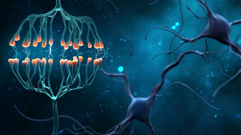
The midbrain, also called the mesencephalon, region of the brain serves important functions in motor activity and movement. 1 A number of structures are located in the mesencephalon including the substantia nigra. 2 Loss of dopamine related neurons within the substantia nigra is the fundamental pathology of Parkinson’s Disease.
The endoplasmic reticulum (ER) is a large, structure that serves many roles in the cell including protein synthesis and proper folding of these protiens.3
Endoplasmic reticulum stress is believed to lead to disruption of protein folding and the aggregation of mis-folded proteins, including the toxic Parkinson’s related protein, α-synuclein, within the neurons. 4
Examination of post-mortem brain tissues has previously revealed a strong association between Parkinson’s disease (PD) pathophysiology and endoplasmic reticulum (ER) stress. Evidence in the literature regarding the circulation of ER stress regulated factors released from neurons provides a rationale for investigating ER stress biomarkers in the blood to aid diagnosis of PD. 5
//////// NGF ////// BDNF GDNF
Parkinson's disease (PD) is a progressive neurodegenerative disorder where dopamine (DA) neurons in the substantia nigra degenerate and die. No cure is available that would stop the dopaminergic decline or restore function of injured neurons in PD. 6 Since no cure for PD exists, there is a need for disease-modifying drugs.
This stimulated research in the direction of nerve growth factors.
Glial cell line-derived neurotrophic factor (GDNF) and related neurturin (NRTN) can protect and repair DA neurons in neurotoxin animal models of PD. 7
These NGF showed promising results in neurotoxin models of PD. 8 9
In the α-synuclein model of PD, a model more closely resembling human disease, both NGF were unable to rescue dopamine producing neurons. 7 8 9
Subsequent clinical trials of these two NGF failed 8 showing only modest effects in phase two studies. 7
Many studies have demonstrated that mesencephalic astrocyte-derived neurotrophic factor (MANF) has been shown to offer protective effects on neurotoxin based models of Parkinson's disease (PD). It still remains unclear whether MANF can rescue dopaminergic (DA) neurons in an α-synuclein model. .8
One α-synuclein model study results showed MANF alleviated progressive neuronal degen- eration and prevented locomotion defects. Indeed, MANF was show to protect DA neurons .8
Furthermore, it was reported that MANF facilitated the removal of misfolded α-synuclein proteins and rescued the function of damaged DA neurons .8
Since α-synuclein model can better mimic the progression of human PD, in another study investigators overexpressed MANF specifically in DA neurons by using an α-synuclein Caenorhabditis elegans (C. elegans) model. 9
Their results showed MANF alleviated progressive neuronal degeneration and prevented locomotion defects. They concluded that within a α-synuclein model, MANF can protect cilia of DA neurons at an early stage, suggested that MANF participated in the whole process of neuronal degeneration. 9
Likewise they found MANF facilitated the removal of misfolded α-synuclein proteins and rescued the function of damaged DA neurons in this model of PD. 9
In addition to its direct protective qualities related to neurons, MANF
demonstrates an ability to modulate levels of other neurotrophic factors, glial cell line-derived neurotrophic factor (GDNF) and brain-derived neurotrophic factor (BDNF), according to previously reported studies . 10
These studies suggest MANF exhibits potential as a neuroprotective agent for PD therapy.
//// ATYPICAL NGF//////
Cerebral dopamine neurotrophic factor (CDNF) and mesencephalic astrocyte-derived neurotrophic factor (MANF) are proteins that have received increasing attention in recent years. Although they are called neurotrophic factors they are drastically different from classical neurotrophic factors in their expression and physiological actions. 11
Neurotrophic factors (NTFs), cerebral DA NTF (CDNF) and mesencephalic astrocyte-derived NTF (MANF) form a novel family of evolutionarily conserved, endoplasmic reticulum (ER) located and secreted NTFs. 7
CDNF and MANF have a unique structure and an unparalleled dual mode of action that differs from other known NTFs. Both protect cells from ER stress, and regulate the unfolded protein response via interacting with chaperons, and CDNF dissolves intracellular α-synuclein aggregates. 7
Mesencephalic astrocyte-derived neurotrophic factor (MANF) and cerebral dopamine neurotrophic factor (CDNF) are endoplasmic reticulum (ER) luminal proteins that confer trophic activities in a wide range of tissues under diverse pathological conditions. Despite initially being classified as neurotrophic factors, neither protein structurally nor functionally resembles bonafide neurotrophic factors. 12
CDNF and MANF are structurally and functionally clearly distinct from the classical, target-derived neurotrophic factors (NTFs) that are solely secreted proteins. In cells, CDNF and MANF localize in the endoplasmic reticulum (ER) and evidence suggests that MANF, and possibly CDNF, is important for the maintenance of ER homeostasis. 13
Despite their misleading names, MANF, together with its closest relative CDNF, form a novel family of unconventional NTF that are both structurally and mechanistically distinct from other growth factors. 14
Neurotrophic factors (NTFs), e.g., glial cell line-derived neurotrophic factor (GDNF) are small, secreted proteins that promote neuron survival during mammalian development and regulate adult neuronal plasticity, and they are studied as potential therapeutic agents for the treatment of neurodegenerative diseases. 6
Different from classical NTFs, CDNF can function both as an extracellular trophic factor and as an intracellular, endoplasmic reticulum (ER) luminal protein that protects neurons and other cell types against ER stress. 6
Despite initially being classified as a neurotrophic factor, MANF has been demonstrated to have restorative and protective effects in many different cell types such as neurons, liver cells, retinal cells, cardiac myocytes, and pancreatic β cells. 15
Mesencephalic astrocyte derived neurotrophic factor (MANF) and cerebral dopamine neurotrophic factor (CDNF) are novel evolutionary conserved trophic factors, which exhibit cytoprotective activity via negative regulation of unfolded protein response (UPR) and inflammation. 10
//////////UNFOLDED PROTEIN RESPONSE ////////
Prolonged ER stress, via the UPR signaling pathways, contributes to the pathogenesis in a number of chronic degenerative diseases, and is an important target for therapeutic modulation. 14
Parkinson’s disease (PD) pathology involves progressive degeneration and death of vulnerable dopamine neurons in the substantia nigra. Extensive axonal arborization and distinct functions make this type of neurons particularly sensitive to homeostatic perturbations, such as protein misfolding and Ca2+ dysregulation. 3
Since ER stress is thought to be one of the pathophysiological mechanisms contributing to the dopaminergic degeneration in PD, CDNF, and its small-molecule derivatives that are under development may provide useful tools for experimental medicine and future therapies for the treatment of PD and other neurodegenerative protein-misfolding diseases. 6
Mesencephalic astrocyte-derived neurotrophic factor (MANF) is a soluble endoplasmic reticulum (ER) luminal protein and its expression and secretion can be induced by ER stress. 15
When misfolded proteins start to accumulate in ER lumen the unfolded protein response (UPR) is activated. UPR is an adaptive signaling machinery aimed at relieving of protein folding load in the ER. 3
When UPR is chronic, it can either boost neurodegeneration and apoptosis or cause neuronal dysfunctions. . 3
MANF and CDNF both can act on unfolded protein response (UPR) genes that modulate the UPR and inflammatory processes . Collectively, this review will highlight MANF and CDNF as broad-acting trophic factors that regulate functions of the endoplasmic reticulum. 12
MANF expression is particularly high in secretory tissues with extensive protein production and thus a high ER protein folding load. 13
Deletion of MANF in mice results in a diabetic phenotype and the activation of unfolded protein response (UPR). 13
By binding to putative plasma membrane receptors, MANF and CDNF promote the survival of DA neurons similarly to conventional NTFs. In animal models of PD, CDNF protects and repairs DA neurons, regulates ER stress, and improves motor function more efficiently than other NTFs. 7
Similarly to the homologous mesencephalic astrocyte-derived neurotrophic factor (MANF), CDNF is able to regulate ER stress-induced unfolded protein response (UPR) signaling and promote protein homeostasis in the ER. 6
CDNF and MANF are localized mainly to the lumen of endoplasmic reticulum (ER) and their primary function appears to be modulation of the unfolded protein response (UPR) pathway 14
////Inflammation
This report discusses the literature supporting a role of Neurotrophic Factors in the regulation of inflammation and regeneration. It focuses, in particular, on the emerging role of mesencephalic astrocyte-derived neurotrophic factor (MANF) and cerebral dopamine neurotrophic factor (CDNF) in the regulation of immune cell function in vivo. Finally, discussing the potential use of these factors to optimize regenerative success in vivo, both within and beyond the nervous system. 16
The excessive activation of the microglia leads to the release of inflammatory factors that contribute to neuronal cell loss and neurodegeneration in Parkinson's Disease (PD). 17
These results suggested that the MANF prevented the dopaminergic neurodegeneration caused by the microglia activation in PD via activation of the AKT/GSK3β-Nrf-2 signaling axis. 17
/////miscellaneous/////////
MANF displays cytoprotective effects in animal models of neurodegenerative diseases. 18
The trophic activities of MANF in tissue repair and regeneration as well as underlying molecular mechanisms propose that MANF might be a promising therapeutic target for tissue repair. 15
Collectively, research findings indicate MANF deficiency affects cell proliferation and suggest a role of MANF in the neurogenesis of the adult brain. 19
Despite multiple reports demonstrating detrimental effect of MANF/CDNF downregulation, little is known about the control of their expression. miRNAs—small non-coding RNAs—are important regulators of gene expression . 10
Here, for the first time researchers demonstrate direct, human-specific, regulation of MANF and CDNF by miRNAs. Identifying that miR-144 controls MANF expression . 10
Under normal conditions, MANF and CDNF are located in the lumen of the endoplasmic reticulum (ER) and their basal secretion from neurons is very low. 11 However in PD patients serum MANF, but not CDNF concentrations were significantly higher in PD patients compared to controls (P < 0.001). 20
Possibly indicating that elevation of MANF is the bodies attempt to mitigate the ER stress caused by misfolded synuclein proteins.
///Therapy
Exogenous MANF and CDNF possess therapeutic properties in several neurological disease models, including Parkinson disease and stroke. Endogenous MANF expression has been shown to be neuroprotective, as well as administration of either CDNF or MANF into the extracellular space. 11
MANF overexpression by adenovirus transduction or addition of MANF into culture media facilitated the growth of longer neurites in RA-treated N2a cells. 21
MANF overexpression facilitates the growth of longer neurites by activating Akt, Erk, mTOR, and P70S6. These findings suggest that MANF positively regulated neurite outgrowth by activating Akt/mTOR and Erk/mTOR signaling pathways. 21
MANF protein could selectively enhance the survival and sprouting of nigral dopaminergic neurons in vitro. Studies have shown that MANF can protect and repair dopaminergic neurons in animal models of PD. 22
Intra-putamenally administered recombinant human CDNF has shown robust neurorestorative effects in a number of small and large animal models of PD, and had a good safety profile in preclinical toxicology studies. 14
In a recent Phase I-II clinical trial CDNF, was found safe and well tolerated. 6
It would seem, based on current research, that supporting and increasing endogenous levels of MANF could prove beneficial in PWP in terms of neuroprotection, if not outright neurogenesis. Exercise despite its many proven benefits in PWP, does not appear to raise MANF levels.
The 4 weeks Mild Intensity Exercise protocol demonstrated positive impact on CDNF (not MANF levels). Whereas Progressive Exercise protocol increased, levels of DA and considerably reduced rotational behavior of rats. The findings of this research confirm positive effects of exercise training in protecting against PD. 23
Cistanche species are herbs used in Traditional Chinese Medicine that show anti-PD potential. 24
Echinacoside (ECH) is a natural phenylethanoid glycoside (PhG) in Cistanche. A large number of studies have shown that ECH has very promising potential in the inhibition of neurodegenerative disease progression. Experimental studies strongly suggest that ECH exhibits a variety of beneficial effects associated with in neuronal function, including protecting mitochondrial function, anti-oxidative stress, anti-inflammatory, anti-endoplasmic reticulum stress (ERS), and regulating autophagy. 25
This study investigated the effects of the Cong Rong Shu Jing (Cistanche CRSJ) compound on endoplasmic reticulum stress in a rat model of Parkinson’s disease (PD). CRSJ administration elevated the expression levels of the neurotrophic factors CDNF and MANF, as well as those of p-PI3K and p-AKT. CRSJ compound can relieve endo
plasmic reticulum stress in PD rats and exerts protective effects in this animal model by increasing expression of neurotrophic factors (MANF) and activation of the PI3K/ AKT pathway.26
Lithium and Piperine (black pepper) appear to support endogenous MANF induction.
The purpose of this study was to determine whether lithium increases MANF expression using cellular and rodent models and, if so, to elucidate the cellular mechanisms of action. Following drug treatment, brain tissue was isolated, and mRNA was extracted from various regions. MANF gene expression was measured. In vitro studies showed lithium-treated cells displayed a significant increase in MANF mRNA expression compared to controls. Similarly, in vivo studies revealed that lithium-treated rats compared to controls had a significant increase in MANF expression in the PFC and striatum. Taken together, these data suggest that lithium's therapeutic mechanism involves the maintenance of ER homeostasis via increased MANF gene expression mediated by the AP-1 transcription factor. 27
Researchers identified several potential candidates that can induce the expression of MANF. Of these inducers, piperine, from black pepper is an agent that potently induces MANF expression. The addition of piperine in both cellular and mouse models of SCA17 alleviated toxicity caused by mutant TBP. Piperine plays its protective role by reducing toxicity caused by the ER stress. 28
The fatty acid DHA may enhance neurogenesis at least in part, through MANF-related mechanisms.
The microglia cell surface receptor (triggering receptor expressed on myeloid cells-2; TREM2) regulates the production of pro- and anti-inflammatory mediators after stroke.
Researchers studied MANF and TREM2 expression after stroke and explored the effects of docosahexaenoic acid (DHA) treatment.
MANF was upregulated in neurons and astrocytes on days 1, 7, and 14 post-stroke.
DHA improved neurobehavioral recovery, attenuated infarct size on days 7 and 14 and increased MANF. MANF and TREM2 protein abundance is robustly increased after stroke and DHA treatment potentiated MANF abundance, decreased TREM2 expression, improved neurobehavioral recovery, reduced infarction, and provided enhanced neuroprotection. 29
……………………References……………….
1. Midbrain | Anatomy & Function | Britannica. britannica.com/science/midb....
2. Learn About the Mesencephalon (Midbrain) Function and Structures. ThoughtCo thoughtco.com/mesencephalon....
3. Kovaleva, V. & Saarma, M. Endoplasmic Reticulum Stress Regulators: New Drug Targets for Parkinson’s Disease. J. Park. Dis. 11, S219–S228 (2021).
4. Ochneva, A. et al. Protein Misfolding and Aggregation in the Brain: Common Pathogenetic Pathways in Neurodegenerative and Mental Disorders. Int. J. Mol. Sci. 23, 14498 (2022).
5. Mnich, K. et al. Endoplasmic Reticulum Stress-Regulated Chaperones as a Serum Biomarker Panel for Parkinson’s Disease. Mol. Neurobiol. (2022) doi:10.1007/s12035-022-03139-0.
6. Lindholm, P. & Saarma, M. Cerebral dopamine neurotrophic factor protects and repairs dopamine neurons by novel mechanism. Mol. Psychiatry 27, 1310–1321 (2022).
7. Voutilainen, M. H., Arumäe, U., Airavaara, M. & Saarma, M. Therapeutic potential of the endoplasmic reticulum located and secreted CDNF/MANF family of neurotrophic factors in Parkinson’s disease. FEBS Lett. 589, 3739–3748 (2015).
8. Zhang, J.-X. et al. MANF Inhibits α-Synuclein Accumulation through Activation of Autophagic Pathways. Oxid. Med. Cell. Longev. 2022, 7925686 (2022).
9. Zhang, Z. et al. MANF protects dopamine neurons and locomotion defects from a human α-synuclein induced Parkinson’s disease model in C. elegans by regulating ER stress and autophagy pathways. Exp. Neurol. 308, 59–71 (2018).
10. Konovalova, J. et al. Human-Specific Regulation of Neurotrophic Factors MANF and CDNF by microRNAs. Int. J. Mol. Sci. 22, 9691 (2021).
11. Albert, K. & Airavaara, M. Neuroprotective and reparative effects of endoplasmic reticulum luminal proteins - mesencephalic astrocyte-derived neurotrophic factor and cerebral dopamine neurotrophic factor. Croat. Med. J. 60, 99–108 (2019).
12. Jӓntti, M. & Harvey, B. K. Trophic activities of endoplasmic reticulum proteins CDNF and MANF. Cell Tissue Res. 382, 83–100 (2020).
13. Lindahl, M., Saarma, M. & Lindholm, P. Unconventional neurotrophic factors CDNF and MANF: Structure, physiological functions and therapeutic potential. Neurobiol. Dis. 97, 90–102 (2017).
14. Eremin, D. V., Ilchibaeva, T. V. & Tsybko, A. S. Cerebral Dopamine Neurotrophic Factor (CDNF): Structure, Functions, and Therapeutic Potential. Biochem. Biokhimiia 86, 852–866 (2021).
15. Deng, H. et al. Emerging trophic activities of mesencephalic astrocyte-derived neurotrophic factor in tissue repair and regeneration. Int. Immunopharmacol. 114, 109598 (2023).
16. Sousa-Victor, P., Jasper, H. & Neves, J. Trophic Factors in Inflammation and Regeneration: The Role of MANF and CDNF. Front. Physiol. 9, 1629 (2018).
17. Zhang, J.-X. et al. Mesencephalic astrocyte-derived neurotrophic factor (MANF) prevents the neuroinflammation induced dopaminergic neurodegeneration. Exp. Gerontol. 171, 112037 (2023).
18. Pakarinen, E., Lindholm, P., Saarma, M. & Lindahl, M. CDNF and MANF regulate ER stress in a tissue-specific manner. Cell. Mol. Life Sci. CMLS 79, 124 (2022).
19. Wang, Y. et al. Deficiency of mesencephalic astrocyte-derived neurotrophic factor affects neurogenesis in mouse brain. Brain Res. Bull. 183, 49–56 (2022).
20. Galli, E. et al. Increased Serum Levels of Mesencephalic Astrocyte-Derived Neurotrophic Factor in Subjects With Parkinson’s Disease. Front. Neurosci. 13, 929 (2019).
21. Wen, W. et al. Mesencephalic Astrocyte-Derived Neurotrophic Factor (MANF) Regulates Neurite Outgrowth Through the Activation of Akt/mTOR and Erk/mTOR Signaling Pathways. Front. Mol. Neurosci. 13, 560020 (2020).
22. Yang, C. & Gao, Y. Mesencephalic astrocyte-derived neurotrophic factor: A treatment option for parkinson’s disease. Front. Biosci. Landmark Ed. 25, 1718–1731 (2020).
23. Fallah Mohammadi, Z., Falah Mohammadi, H. & Patel, D. I. Comparing the effects of progressive and mild intensity treadmill running protocols on neuroprotection of parkinsonian rats. Life Sci. 229, 219–224 (2019).
24. Lei, H. et al. Herba Cistanche (Rou Cong Rong): A Review of Its Phytochemistry and Pharmacology. Chem. Pharm. Bull. (Tokyo) 68, 694–712 (2020).
25. Li, J., Yu, H., Yang, C., Ma, T. & Dai, Y. Therapeutic Potential and Molecular Mechanisms of Echinacoside in Neurodegenerative Diseases. Front. Pharmacol. 13, 841110 (2022).
26. Lin, Y. et al. Mechanisms of Cong Rong Shu Jing Compound Effects on Endoplasmic Reticulum Stress in a Rat Model of Parkinson’s Disease. Evid.-Based Complement. Altern. Med. ECAM 2020, 1818307 (2020).
27. Abu-Hijleh, F. A. et al. Novel mechanism of action for the mood stabilizer lithium. Bipolar Disord. 23, 76–83 (2021).
28. Guo, J. et al. Piperine ameliorates SCA17 neuropathology by reducing ER stress. Mol. Neurodegener. 13, 4 (2018).
29. Belayev, L. et al. DHA modulates MANF and TREM2 abundance, enhances neurogenesis, reduces infarct size, and improves neurological function after experimental ischemic stroke. CNS Neurosci. Ther. 26, 1155–1167 (2020).