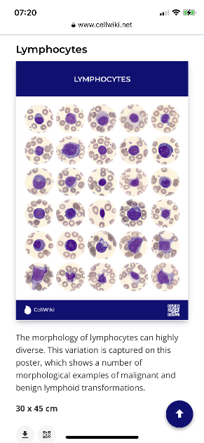
This poster from CellWiki show just how variable the morphology of lymphocytes can be. This poster includes both normal and malignant lymphoid cells. Of course, cells don’t read the books or poster and there will be others not shown here.
It can be extremely difficult to positively identify a cell by morphology alone and that is why flow cytometry with a range of anti CD reagents is used to ‘type’ them. The combination of those results uniquely identifies the type of cell.
Jackie
Written by
Jm954
Administrator
To view profiles and participate in discussions please or .
The stain is one called May Grunwald Geimsa, one of the Romanowsky stain family and on blood smears produces shades of pink to dark blue depending on the pH
Romanowsky stains are neutral stains composed of a mixture of oxidized methylene blue (azure) dyes and Eosin Y. The azures are basic dyes that bind acid nuclei and result in a blue to purple color. The acid dye, eosin, is attracted to the alkaline cytoplasm, producing red coloration.
Thanks , Jackie. If gives me more clarity to the need for the shotgun vs the pistol approach of the early chemo drugs and the later novel agents like ibrutinib at the highest dosage.
Now, I understand why pathology reports have used the term *rare* when describing *neutrophils* - The poster shows the dilemma for cells in general - thank you.
neutrophils are only rare in the relative sense because there are so many lymphs. Rare is a bit of a daft description because the numbers tell the story of how many there are.
You would not see all this lymph morphology in one person’s blood. This is just showing how diverse they can be in different pathologies.
Thank you. To be clear - I do understand the chart is not representative of what you would see in one person’s blood. The term - *rare* anything - for calcifications and or neutrophils always confused me. It was used in 2 separate pathology reports. I asked if it was the *number* present or were they deformed? (wrong term - help me please) in some way. The response in one was - *bottom line - get it out now* - for the DCIS and
*rare*occurrence for the *rare* neutrophils noted in the colonoscopy bx. Path report. It was a bx of the illocecal valve.
Goodness - and here I was thinking fully formed or immature. I suppose I was aware that there must be other forms of “wrong” for acute leukaemia or multiple myeloma for instance but that chart is illuminating. I always did have respect for the people reading the blood tests ... (Which one(s) are “ours” - or is that not simple either?)
I've had unusual lymphocytes for years - atypical, reactive, abnormal, and variant lymphocytes. Based on PubMed searches, those terms are not well defined, and there is overlap in meaning. Much depends on the pathologist reviewing the slide or the particular model of automated blood analyzer and how they describe the size, shape, and nucleus.
Automated analyzer reportas attempt to "flag" unusual cell types. Sometimes their algorithms are not so successful. Anything flagged is supposed to be reviewed manually. But manual reviews are also not so consistent.
Cell Cytometry: Review and Perspective on Biotechnological Advances
Published online 2019 Jun 18.
Pathologists may be swayed by the flagging of the automated analyzer.
Manual differentials are a skill, and there's much room for subjectivity, especially with odd cell morphology. Some labs do a wedge smear, which may not distribute all types of cells consistently.
Rodak's Hematology Clinical Principles and Applications, PART III, Laboratory Evaluation of Blood Cells, CHAPTER 16, Examination of the peripheral blood film and correlation with the complete blood count
Some labs use a hemocytometer - a special type of microscope slide that makes counting easier:
Again, there is room for subjectivity for unusual cell morphology. Either method uses only about 100 cells to calculate the differential, where an automated analyzer uses thousands. Training for lab technicians focuses on typical, often normal blood counts. Lab techs ask pathologists to review unusual results, but staffing stresses makes this all more hasty than efficient.
I think a recent model automated counter with 5 or more colored lasers produces pretty reliable results, even for unusual morphology.
The issue I have with all this is that repeated results with unusual morphology indicates something. But since most people get tested only once or twice a year, and since specialists are used to patients with that sort of frequency, they will not try to chase down a cause, because it could have been a temporary thing. Electronic medical records often have no "field" for unusual cell types - the whole record must be read. Specialists instead look for other symptoms to correlate with the results. But the symptoms of a chronic infection or inflammation that can cause unusual lymphocytes overlaps with the symptoms of CLL itself. Lack of effective antivirals discourages virus testing. So we're stuck. The terminology used by the pathologist - abnormal, vs. atypical vs. reactive - along with any text descriptions of size and nucleus can really help. But not all pathologists are so verbose or careful with descriptions.
I also really wish that test results would list the model and version of automated analyzers as a necessary consumer and medical quality control. Some hospital labs keep old, 3 color analyzers for years. A knowledgeable specialist could then request a test at a better lab. Research labs often have the best equipment that even photographs each cell for review by others.
Content on HealthUnlocked does not replace the relationship between you and doctors or other healthcare professionals nor the advice you receive from them.
Never delay seeking advice or dialling emergency services because of something that you have read on HealthUnlocked.