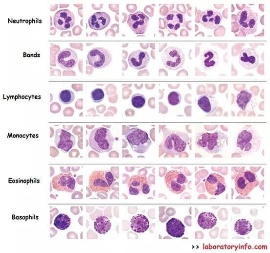
I thought some members might like to see these images of what the various 'normal' white blood cells look like. As you can see there is a variability of morphology within each type of cell. Bands are a slightly immature form of neutrophil and as it matures the nucleus becomes more segmented into lobes like the pictures in the neutrophils line.
Lymphocytes are either large or small and the first four from the left in this photograph are small lymphs and the two on the right are large lymphs. Large lymphs are often T cells and have more cytoplasm, often with a few granules in it. Activated T cells which are generally large with expansive, dark cytoplasm are often noted as atypical lymphs on blood reports and can be indicative of infection.
Monocytes are extremely variable and the nucleus may be indented or not and the cytoplasm may have vacuoles in it, or not. Large lymphs and monocytes can sometimes be hard to distinguish but as a rule of thumb, monocytes are bigger (but not always) and large lymphs may have a few pink granules (but may not).
The pink/orange discs in the back ground are red cells and that's a whole new morphology game  . Platelets are the smallest cells in the blood and they are actually just fragments that have broken off from 'buds' that form on the precursor megakaryocyte cells in the bone marrow. They have no nucleus and can vary hugely in size from tiny specs to the size of a lymphocyte. If you look at the lymphocyte's line then the fourth from the left has two platelets at approx 10/11 o'clock and in the monocyte's line, the second from the left has a larger platelet at about 3 o'clock.
Importantly, all these cells were photographed at the same magnification so you can actually see the relative sizes.
Morphology is fascinating because the cells don't read the morphology books and just do their own thing! I hope you've found this interesting.
Jackie
