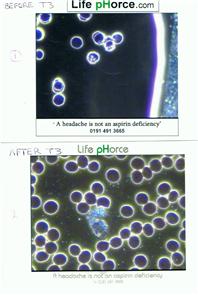
Live blood analysis photographs. Quite serious before. Excellent After!!
Photograph of my blood cells before and after T... - Thyroid UK
Photograph of my blood cells before and after T3 treatment.
Read more about...
10 Replies
•
hi wildflower,
This is very interesting. Can you explain in a bit of detail for us please? For example, what is it showing in the blood? What was the treatment which made the improvement? The background to the tests, etc. Many thanks!
Mary
Sorry just re-read title it says after T3 treatment! Were you being treated with anything before?
in reply to MaryMary
Yes, I was on levothyroxine since April 2005
Kept being ill and they kept putting my dose up, ended up on 150mg a day.
(Photo1) Taken later in the year 2005
Stopped Levothyroxine 3/8/2010 as I was very toxic, and not converting.
Started T3-only treatment 4 weeks later.
(Photo 2) 19/5/2011. 9 months after stopping levothyroxine and starting T3-only treatment.
After only 2 weeks on T3 my severe pain had gone.
After 2 months my Creatinine Kinase mm test ( which is the muscle enzyme test, shows that my muscles were breaking down, hence the severe pain).
My CKmm which had been raised for over 10 years was back to normal.
Cholesterol which was 9 was also back down to normal.
Hope you can understand this!
Brain foggy at the mo but how fascinating thanks for posting! ")
Hi
This is brilliant and something that really interests me, health wise and professionally. Could I ask how you came to have this done or where you had this done? Thanks
D
That's REALLY interesting. Who did the blood tests like this? Who took the photographs? Is this something that could, possibly, be a future test that could demonstrate or prove (via, no doubt, some sort of study) that T3 actually DOES work that the doctors would believe? I think that's absolutely fantastic and I want to hear and see more!
in reply to parafluie
Test done by :- Live Blood Analysis; you can actually see on screen what your blood cells are doing. A photo is taken from the screen. As I said they cannot diagnose, but they can tell you what you are deficient in.
As T3 gives oxygen to the cell, I can only guess what happened to me. The practitioner did say " what have you been taking, all the nasties have gone".
You can see the nasty bits on the cells edges on the first photo. He knew what
they were called.
That rather large thing in the second photo is a white blood cell, cleaning up.
Amazing. So the practitioner does the analysis as opposed to a Lab. Dunno if there's anything like that in USA. Frankly, if changes like that are clear for many people, I'd like to see a clinical study. It's amazing. Thanks for sharing.
Not what you're looking for?
You may also like...
Blood results after adding T3
I am on 75mcg of levothyroxine & 6 weeks ago I added 5mcg of Liothyronine & 2 weeks ago I went up...
Blood test after adding t3
If it takes 6-8 weeks for levo to show full effect on blood tests when the half life is 7 days...
Blood work before of after tirosint-what’s the difference?
Hello everyone,
I need your advice. My doctor in the USA said that it doesn’t matter when you do...
Anyone know of a good before and after thyroid treatment comparison template?
I will be seeing my GP in ten days and if I continue at the level of improvement I am experiencing...
T3 and Blood Pressure
Hi there...my case should be well known by now and is not uncommon.......undiagnosed and then under...