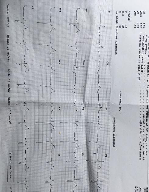
The following ECG was taken today prior to a planned angiogram following chest pains (which didn't take place owing to an issue with sedation). It apparently shows left bundle branch block, left axial deviation and inverted T waves. Does anyone know what the significance of these three is? Or how marked these are on the ECG? I'm guessing its not good.
Help needed interpreting ECG. - Atrial Fibrillati...
Help needed interpreting ECG.

I would not get overly stressed about this until you speak to an ep. One of my EKGs showed left bundle branch block while on Flecainide and my EP did not make a big deal of it.
Jim
Thanks for the reply.
We really can't advise on such matters as we are not medically trained but I do know that inverted T waves are ectopic beats so benign.
Inverted T waves are normal in children and some adults.
If your T waves have historically been negative then it is perfectly normal for you. Mine are normally inverted.
Only if this T wave changes to flat or positive would I become interested in it.
On some ECG leads if the R wave is net negative then the T wave would also be negative.
LBBB is normal for people with Bradicardia. The EP would not be concerned with LBBB.
Thanks for the reply. I have bradycardia-at least after taking Sotolol-so hopefully that explains it. Part of the problem I'm having at the moment is that the hospital doctor carrying out the angiogram is not coordinating very well with the consultant and is reluctant to comment on anything for fear of treading on his toes.
Oh dear Samazeulh
It looks as if your heart is under stress.
Better to leave things.
Have you had an ECHO of your heart?
On Metoprolol my heart showed under stress. Finally I was changed under a Cardiac Specialist then left again UNCONTROLLED. CONTROL is so important. As you know the CCB Diltiazem keeps me controlled.
Now the 'talk' is AF comes with inflammation anywhere in your body.
Under your Specialist an ECG is done and he/she talks about it. Has no one explained your ECG?
Pity
I always ask for a copy as well.
There is an ECG machine in our clinic.
Take care.
I cant have anything done like Cardiconversion or Ablation because ECHO shows an enlarged heart when they scanned me last Feb 21.
The latest secialist found that I had a soft systolic murmur (often called 'innocent murmur'.
cheers JOY. 73. (NZ)
I also have a "wide QRS" and LBBB, but I haven't seen inverted T waves on mine, yet I have read this can happen with branch block. I, too, have been told that the LBBB rarely comes to much, but the wide QRS does seem to be responsible for my having lots of ectopics and chest symptoms and a need to breathe in rather deeply at times. It was like that last night, for example, accompanied by mild tachycardia but no AF and even a bisoprolol didn't help much.
Your GP, if not your specialist, needs to have a look to advise more. If you have any physical changes ion your heart shown by an echo scan, for example, they will know this and the ECG consequences of it.
Steve
Thanks for the reply. They have forwarded the ECG tontge consultant who will hopefully advise on it soon. The left axial deviation I have had for years (even before getting AF). Not sure if it has got worse.
You mean back block? From what I’ve read, it usually becomes a complete “block” but the heart uses other pathways. These are a fraction slower, creating the “wide qrs“.
It’s hard to find anything on the internet about symptoms, most comments are that it is symptomless, but some do mention various other symptoms.
Steve
I’m not familiar with the term “back block”; so far as I know it hasn’t been used in relation to the ECG. The doctor mentioned the possibility of myocarditis but said he couldn’t say what the cause of the abnormal ECG is.
Oh dear - you can guess I’m tired. Sorry - it was the autocorrect on this phone!!! I meant branch block (left or right). A cardiac stress MRI is the gold standard scan I gather.
Steve
I would advise speaking to your doctor about the ECG results, they are medically trained to provide an overall assessment and arrange for any necessary follow up's or treatment options to be followed.
Thanks.
Hi hon, instead of worrying can’t you ask your doctor or a nurse? I think those high peak ones that are hanging upside down are the inverted because for the first time I had one NSR and the tall peaks are very uniform but going opposite yours. Prior to my last one they were all over the place and all different shapes.
As far as the angiogram, I was given some type of a cocktail through my IV that just made me silly more than anything I think. They went through my wrist and it was very uneventful. Maybe there is something similar they can do for you. I recently had an echocardiogram which is very non-invasive and nothing needed to have it done. I’m just not sure what needs to be diagnosed with you. Usually asking your doctor or the doctors office is your best bet. The PN at my doctors office is incredible and I have no problem dealing with her most of the time because my doctor is so busy performing procedures.
Thanks for the reply. I'm being investigated for suspected angina. I asked the doctor who was to carry out the procedure what the significance of the abnormal ECG was and he simply said it could be "a number of different things" and said I would need to speak to the cardiologist who referred me. I rang his secretary but he was not available. She has emailed him, but I am not expecting a response any time soon.
In my opinion you have anterior ischemia of your heart , probably your left anterior descending coronary artery has some blockage. Only an angiogram can decide of the next steps to follow, probably a stent or two . In the mean time I hope you have a medical treatment to keep you going . In US, ecg like that is an emergency, which means an angiogram on the fly. I am so surprised and disappointed that things turned out that way for you. You would need a plumber here and not an electrician cardiologist, who specializes in putting a stent during angiogram procedure.
Thanks for the reply.