Radiation, Focal or Removal? - Prostate Cancer N...

Prostate Cancer Network

4,955 members3,087 posts

Radiation, Focal or Removal?

Swampfox profile image
30 Replies

I was just diagnosed last week. Here is the layout of the biopsy results. 13 cores with a combination of 3+3=6 and 3+4=7. I meet with the doctor on Monday, He has already ordered a PRolaris Genomic test. Should get that back in 4 weeks. I am going to look at all options before making any decisions. I am 58 and healthy. Question: Has anyone with similar biopsy results had focal therapy?

Thanks,

Marc

Final Diagnosis

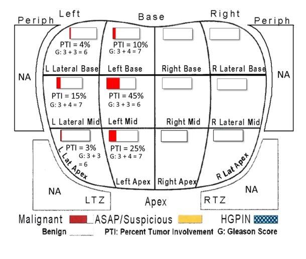
A. Prostate, left lateral base, biopsy: Adenocarcinoma of prostate, Gleason score 3 + 3 = 6, Grade Group 1, involving one core (0.5 mm, <5% of tissue present)

B. Prostate, left lateral mid, biopsy: Adenocarcinoma of prostate, Gleason Score 3 + 4 = 7 (pattern 4 = 5%), Grade Group 2, involving one core (2 mm, 15% of tissue present)

C. Prostate, left lateral apex, biopsy: Adenocarcinoma of prostate, Gleason score 3 + 3 = 6, Grade Group 1, involving one core (<0.5 mm, <5% of tissue present)

D. Prostate, left base, biopsy: Adenocarcinoma of prostate, Gleason Score 3 + 4 = 7 (pattern 4 = 5%), Grade Group 2, discontinuously involving, one core (1.5 mm, 10% of tissue present)

E. Prostate, left mid, biopsy: Adenocarcinoma of prostate, Gleason score 3 + 3 = 6, Grade Group 1, involving one core (7 mm, 45% of tissue present) Perineural invasion present

F. Prostate, left apex, biopsy: Adenocarcinoma of prostate, Gleason Score 3 + 4 = 7 (pattern 4 = 10%), Grade Group 2, involving one core (2.5mm, 25% of tissue present) Perineural invasion present

G. Prostate, right base, biopsy: Benign prostate tissue

H. Prostate, right mid, biopsy: Benign prostate tissue

I. Prostate, right apex, biopsy: Benign prostate tissue

J. Prostate, right lateral base, biopsy: Benign prostate tissue

K. Prostate, right lateral mid, biopsy: High grade prostatic intraepithelial neoplasia (HGPIN)

L. Prostate, right lateral apex, biopsy: Benign prostate tissue

M. Prostate, PI-RADS 4 left apex PZ, MRI-guided biopsy:

Adenocarcinoma of prostate, Gleason Score 3 + 4 = 7 (pattern 4 = 10%), Grade Group 2, involving four cores (19mm, 35% of tissue present) Perineural invasion present

Written by
Swampfox profile image
Swampfox
To view profiles and participate in discussions please or .
Read more about...
30 Replies
Tall_Allen profile image
Tall_Allen

Focal therapy doesn't work. And there is a lot of hype from unscrupulous urologists. Read this:

prostatecancer.news/2016/12...

It may not change your diagnosis, but it's probably a good idea to get a second opinion from Epstein's lab at Johns Hopkins:

pathology.jhu.edu/patient-c...

Swampfox profile image
Swampfox in reply to Tall_Allen

Thank you. The second opinion at Johns Hopkins is in progress. Thanks for the article link

in reply to Swampfox

Definitely do your own research. ncbi.nlm.nih.gov/pmc/articl... this is a later article on FT. Had I known about it, I think I would have tried this treatment. RP is ghastly.

Swampfox profile image
Swampfox in reply to

Thank you

dentaltwin profile image
dentaltwin in reply to Swampfox

Remember that each man's experience is different. My RP was not "ghastly". You have time to make your decision--take it.

dlste353 profile image
dlste353 in reply to dentaltwin

I did not find RP ghastly either.

GeoffNoLongerAS profile image
GeoffNoLongerAS in reply to Swampfox

Nor was mine. Each therapy has its positives and drawbacks. Learn about each therapy, the outcomes and risks.

It seems from the diagram you provided that though not terribly aggressive, tumors were found in a number of areas. Some questions I would have (rhetorical) is did you have an MRI? Were there suspicious lesions indicated by the MRI elsewhere? Would it make better sense to treat the entire prostate (RP or radiation)?

I hope all goes well for you in whatever treatment you choose.

Swampfox profile image
Swampfox in reply to GeoffNoLongerAS

Yes. I had an MRI that detected a lesion. They used the MRI to target the lesion during biopsy. Thanks for your input

Swampfox profile image
Swampfox in reply to Swampfox

Lesion was in the prostate, I should have said. I am going to ask for a PSMA Petscan to make sure nothing jumped the prostate

MauiJef profile image
MauiJef

Sorry to hear you've joined our club, you'll get tons of advice and suggestions in here. It's been a very good source for me. I'm still deciding on my treatment path but I have read several books that have helped me along the way.

Dr. Patrick Walsh's Guide to Surviving Prostate Cancer

I'm currently reading this one and its very informative

The Key to Prostate Cancer: 30 Experts Explain 15 Stages of Prostate Cancer from

Dr. Scholz

Good luck in your journey

Swampfox profile image
Swampfox

Thank you. I have been watching the videos from Dr Scholz also and they have been very helpful. Trying to get all the info that I can to make the best decisions

Lightjunkie profile image
Lightjunkie

Sorry you joined the club! Sounds like you are on mark with your last comment, “trying to get all the info I can to make the best decisions.” There’s so much to learn, it feels like there’s no time, yet, somehow I keep learning and turned on to so much information and wisdom here. Good luck!

Bigmls7890 profile image
Bigmls7890

Get PSMA pet scan make sure gland contained get an opinion for FLA and or Tulsa from Dr Eric Walser , UTMB, Houston, TX.

Swampfox profile image
Swampfox in reply to Bigmls7890

Thank you

tsim profile image
tsim

I'm going to ask a couple of simple questions since there have been some real nightmare stories on here lately about complete and really negligent diagnosis and maltreatment.

Other than the PNI did the MR show any nodal involvement or any other involvement outside the prostate?

Have you had a bone scan?

Was your biopsy done by a urologist or an oncologist?

Relax, you've got time to decide treatment.

Swampfox profile image
Swampfox in reply to tsim

Biopsy was done by an oncologist. I had an MRI before showing the lesion, but no evidence it has left the prostate. No nodal involvement

tsim profile image
tsim in reply to Swampfox

That's good, I'll assume you've had or have scheduled a bone scan. Your diagnosis was similar to mine with the exception of a small tumor in my left seminal vesicle. I chose HDR brachytherapy combined with 18 months ADT and that worked out quite well. PSA undetectable after 5 years and almost no side effects. Take your time and ask as many questions as you need to so you can understand the options. And, as TA said, focal not effective.

Swampfox profile image
Swampfox in reply to tsim

Thank you!

cscmetsfacil profile image
cscmetsfacil

No diagnosis of cancer is "good", obviously, but yours could be a lot worse - that's the good news. The "bad" news is that you have a lot of options to work through. Your doctor is doing the right thing getting the genomic testing. You might look at a Decipher test as well. The goal is to understand whether you have any genomic conditions that could move your "favorable" GL7 into the "unfavorable" category. You also want to see if there is any cancer close to the capsule and any found in nerve tissue. Looks like the larger burden of cancer is toward the center of the gland. In any event, there is an emerging school of thought that there is no reason to opt for surgery (which is a seriously invasive procedure) when there are new, precise methods of ablative radiation that will spare your nerves and have excellent results with many fewer side effects. I would certainly check out either HDRT or SBRT alternatives before going with surgery. If you want to chat, I facilitate PCa support groups at the Cancer Support Community in Walnut Creek, CA, and can be reached at vortiz@cancersupport.net Wishing you all the best.

Swampfox profile image
Swampfox in reply to cscmetsfacil

I do have "encapsular abutment" as found in the MRI, so it is close to the edge, but has not left the prostate, as far as we know. From what I have found, radiation therapy has comparable success rates to surgery, assuming the doctor deems me a candidate for radiation. Thank you for your response and input

CarverD profile image
CarverD

If your diagnosis remains the same after the JH opinion you have just about every treatment option available to you for favorable PCA. I narrowed my decision by looking at the side effects of each, both short and long term. After 9 months of research and discussions with 5 doctors I opted for HDBT. Three years out and I'm still pretty happy with the results.

Swampfox profile image
Swampfox in reply to CarverD

Glad to hear it went well for you. I am following the same road as you in regard to taking it slow and reviewing all of my options.

Piano777 profile image
Piano777

First, thanks for the great visual of your biopsy result. It's a nice model for seeing results. I wish official biopsy results were published that way.

There is a very recent excellent summary on focal treatments from AnCan that outlines the issues, both pro and con.

youtu.be/Swn-ZoKGjz4?si=MiF...

I had NanoKnife about 1.5 years ago on the left peripheral 4+3 with zero side effects and have been cancer free in-field. However, I now have PCa on the right peripheral 3+4. It's one issue why focal may not be right. I'm investigating radiation as my next step. You may want to look into MRgSBRT (MRI guided SBRT). In a clinical trial it had significantly less adverse events than traditional SBRT.

ascopost.com/news/january-2...

Good luck

Swampfox profile image
Swampfox in reply to Piano777

Thank you. Great info

groundhogy profile image
groundhogy

Here is a good website to compare odds of cure for the major treatment paths. You have to determine your stage, low risk, intermediate, or high risk (risk of recurrence). So if you are intermediate, pull up the intermediate chart and you can see the odds of 10-20 yr survival, etc. based on the treatment you pick.

prostatecancerfree.org/comp...

It is best viewed on computer or just print it on paper. Not so viewable on phone.

To make the graphs easier to read, i drew a dot on the endpoints of the elipses, and then drew a line through the dots. This turns the elipses into lines.

Also be aware the the graphs don’t show any salvage radiation benefit. This would boost the surgery odds up a bit.

Also beware, this is a very dysfunctional industry from my view. Loads of bad info mixed in with the good info. Same with the docs. Some of them are more dangerous than the cancer.

Swampfox profile image
Swampfox in reply to groundhogy

Thank you for sharing this info. This is great

NotDFL profile image
NotDFL

I will repeat what some have written. In addition to the loci identified as cancerous, there are likely to be others (tiny ones) that can't be detected by current methods. Hence, a focal treatment may have a good short-term outcome, but it's pretty likely that more lesions will pop up down the line. Therefore, I recommend a therapy that covers all of the prostate, whether it will be brachy, SBRT or IMRT. Removal is another option, of course.

Swampfox profile image
Swampfox in reply to NotDFL

Thank you. I am definitely going to look at all of my options

the thing about radiation, to me, is it does not involve poking and tearing at a cancerous piece of tissue, which to me, has the possibility of breaking a microscopic piece off. of course the doctors will say this doesn't happen.

billy1950 profile image
billy1950

Hi,

My diagnosis was gleason 3+3 with PSA 6.9…

In 2004-5 ( age 54) i was treated with EBRT and went through it well. No SE except mild fatigue in the 5th to 8th week. RT has improved and evolved since then…

All of us with Pca have had different experiences …Youtube videos and information are so much better today. You seem to have done a lot of research already…Take time to decide in building your confidence before making your choice…My best!

You may also like...

Uro and Surgeon Recommending Focal Cryoablation

appreciated. E. Prostate, left mid, needle core biopsy: - Prostatic adenocarcinoma,...

newly diagnosed Gleason 7 3+4

Prostate, left base; needle biopsy: - Prostatic adenocarcinoma, Gleason grade 3+4=7 (Grade...

Biopsy Question: Suspicious for focal intraductal carcinoma

Cancer Grading: Primary Gleason grade: 4 Secondary Gleason grade: 3 Total Gleason score: 7...

Gleason score change from 3+4 to 3+3 with perineural invasion noted under Active Surveillance

A: Benign prostatic tissue. B. RPZ PL: PROSTATIC ADENOCARCINOMA, Gleason score 3+3=6 (Grade Group 1)

New Diagnosis - feeling overwhelmed

A biopsy of 16 cores showed benign tissue in the Left Lateral Base, Left Base, Right Base, Right...